Biotin- a water-soluble Vitamin
Biotin is a water-soluble vitamin that functions as a coenzyme for five mammalian carboxylases: pyruvate carboxylase (EC 6.4.1.1), propionyl-CoA carboxylase (EC 6.4.1.3), methylcrotonyl-CoA carboxylase (EC 6.4.1.4), and both isoforms of acetyl-CoA carboxylase (EC 6.4.1.2). Biotin, cis-hexahydro-2-oxo-1H-thieno[3,4-d]-imidazoline-4-valeric acid, is a small molecule with the molecular weight of 244.3 dalton. Other synonyms for biotin are D-Biotin, Bios II, Coenzyme R, Vitamin B 7, and Vitamin H.
Biotin is a colorless crystalline powder with the chemical formula C10H16N2O3S. It is part of the vitamin B2 complex and is essential for all mammals including humans. Vitamin B2 was found to be a complex of several chemically unrelated heat-stable factors, including niacin, biotin, and pantothenic acid. Furthermore, it is a cofactor for many enzymes in the body and is found in large quantities in liver, egg yolk, milk, and yeast.
.jpg)
Figure 1: Structural models for biotin. (A) Chemical structure; (B) Stick model of the energy minimized structure; (C) Structure of biotin as observed in a crystal of a deglycosylated avidin in complex with biotin published 1993 by Livnah et al. [2AVI]; (D) Space filling model of biotin.
Biotin was discovered during nutritional experiments that revealed it as a factor in many foods. Biotin was found to be capable of curing scaly dermatitis, hair loss, and neurologic signs induced in rats when fed dried egg whites. Deficiency in biotin causes dermatitis and loss of hair. Biotin is a water-soluble, coenzyme present in small amounts in every living being. It occurs mainly bound to proteins or polypeptides. However, biotin is abundant in liver, kidney, pancreas, yeast, and milk. In humans biotin is a coenzyme for five carboxylases: propionyl-CoA carboxylase, methylcrotonyl-CoA carboxylase, pyruvate carboxylase, and 2 forms of acetyl-CoA carboxylase. This fact makes biotin essential for amino acid catabolism, gluconeogenesis, and fatty acid metabolism.
The enzyme biotin holocarboxylase synthetase (EC 6.3.4.10) catalyzes the attachment of biotin to the apocarboxylases by an amide bond to a specific lysine residue–producing holocarboxylases. Biotinylated proteins are normally turned over by proteolytic degradation to biocytin (biotinyl-lysine) and biotinylated oligopeptides that are subsequently cleaved by biotinidase (EC 3.5.1.12). These reactions recycle biotin. In addition, biotin is found covalently attached to histones and appears to be necessary for gene stability, repression of transposable elements and some genes.
Biotin contains three chiral centers and has eight stereoisomers. However, only one is active. The active form is the dextrorotatory (+)-biotin. Biotin is synthesized from L-alanine and pimelolyl-CoA via a condensation reaction to form 8-amino-7-oxononaoate (AON). Four enzymes are needed for the synthesis. A synthase catalyzes step one, and a transaminase catalyzes step b. The coenzyme pyridoxal phosphate (PLP) is needed as well. The final step is the insertion of a sulfur atom from an iron-sulfur center.
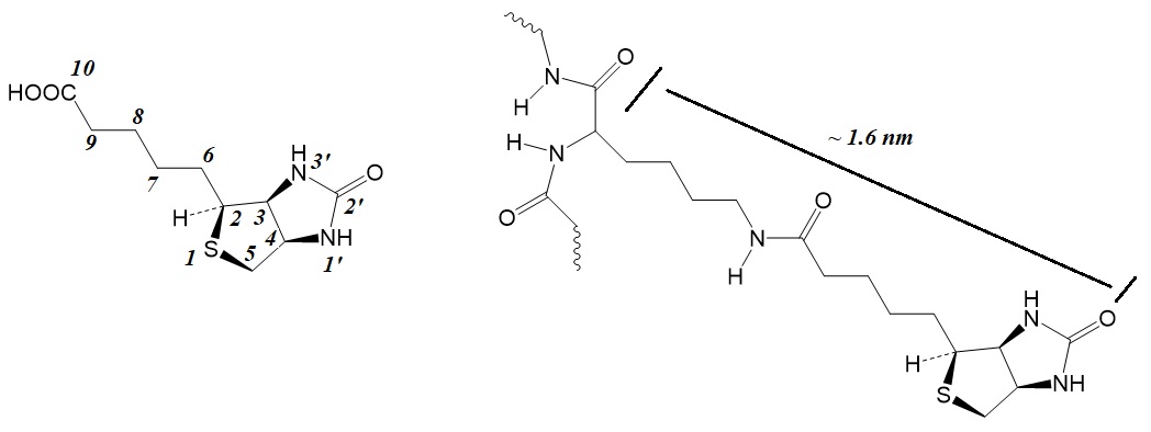
Figure 2: Structure of biotin and biotin conjugated to a protein as a carrier. In cells, biotin is covalently bonded to proteins as a double ring attached to a ~1.6-nm flexible arm. The enzymatic ATP-dependent reaction of pyruvate with bicarbonate ion to form oxaloacetate requires biotin. Other biotin-requiring enzymes are known to act on acetyl-CoA, propionyl-CoA, and beta-methylcrotonyl-CoA, using HCO3- for the addition of carboxyl groups.
Enzymes containing bound biotin required for the catalysis of beta carboxylation reactions include acetyl-CoA carboxylase, propionyl-CoA carboxylase, pyruvate carboxylase, and β-methylcrotonyl-CoA carboxylase (δ carboxylation). The carboxyltransferase of Propionobacterium catalyzes the carboxyl group transfer without cleavage of ATP. Oxaloacetate decarboxylase, methylmalonyl-CoA decarboxylase and glutaconyl-CoA decarboxylase are biotin-dependent sodium ion pumps. Malonate decarboxylase, and urea carboxylase are also dependent on biotin.
Normal dietary intake of foods usually supplies enough biotin to prevent a biotin deficiency. However, deficiency can be caused by eating too many raw egg whites for a prolonged time period. Egg whites contain the protein avidin in large amounts. This protein can bind biotin strongly and prevent it from being ingested. Biotin in the body is regulated by dietary intake, the biotin transporters monocarboxylate transporter 1 and sodium-dependent multivitamin transporter, peptidyl hydrolase biotinidase (BTD), and the protein ligase holocarboxylase synthetase. Inhibiting any of these enzymes can cause a biotin deficiency.
Biotin Deficiencies
Nutritional biotin deficiency and inherited enzymatic deficiencies of the biotin-dependent carboxylases cause abnormally increased urinary excretion of characteristic organic acids.
Structures of observed metabolites
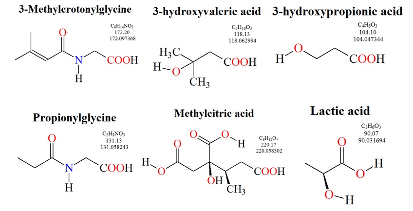
Figure 3: Structures of metabolites observed in urine for biotin deficiencies.
Biotin as a coenzyme is a catalyst for carboxylation reactions. One example is the reaction catalyzed by propionyl-coenzyme A (CoA) carboxylase illustrated in figure 3.
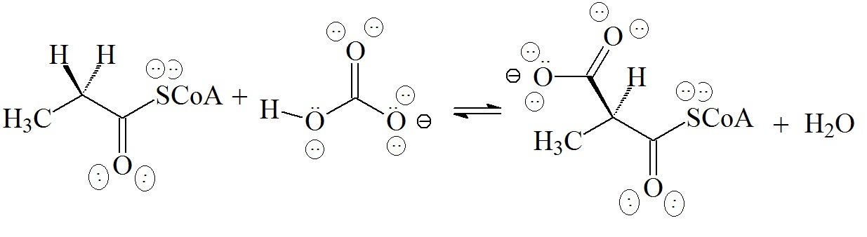
Figure 4: Carboxylation reaction catalyzed by propionyl-CoA carboxylase.
In the reaction catalyzed by propionyl-CoA carboxylase, acidic hydrogen on carbon is removed as a proton and is replaced by the electrophilic acyl carbon of bicarbonate in an acyl exchange reaction. Here, water is the formal leaving group. The energy-yielding reaction for biotin catalyzed carboxylation reactions usually is the hydrolysis of MgATP to MgADP which is coupled to the carboxylation reaction. Biotin is covalently attached to the active site of an enzyme by an amide between the carboxylate of the coenzyme and a lysine from the protein.
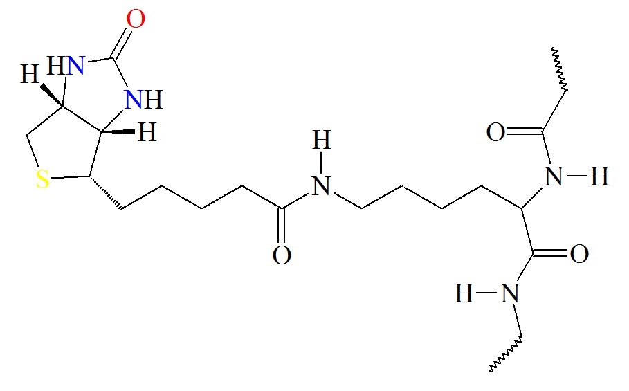
Figure 5: Biotin covalently attached to the active site of an enzyme. The biotin moiety is attached by an amide between the carboxylate of the coenzyme and a lysine from the protein.
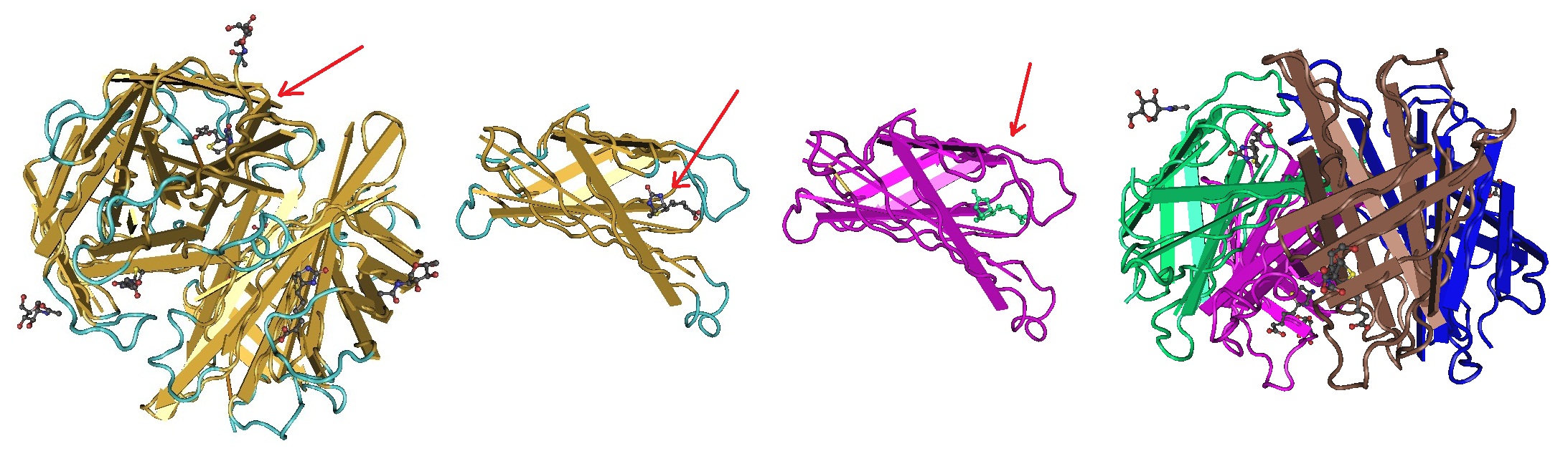
Figure 6: Biotin-Avidin Complex. The crystal structures of a deglycosylated form of the egg-white glycoprotein avidin and the avidin-biotin complex have been determined to 2.6 and 3.0 Å by Livnah et al. in 1993. The structures revealed the amino acid residues critical for the stabilization of the tetrameric protein complex assembly as well as for the very tight binding of biotin. Each avidin monomer folds into a eight-stranded antiparallel beta-barrel structure. This structure is quite similar to that of the genetically distinct bacterial analog streptavidin. Similar to streptavidin, binding of biotin involves a highly stabilized network of polar and hydrophobic interactions. Different views of the tetramer and monomer are illustrated. The red arrow points to the biotin binding pocket. The avidin-biotin interaction is the strongest known noncovalent recognition between protein and ligand (kd = 10-15 M). The bonds between biotin and avidin are rapidly formed. The avidin-biotin complex is very stabile in a wide range of conditions. Biotin binds to avidin at room temperature in the presence of detergents such as SDS, Tween 20, Tween 40, and Triton X-100.
The valeric acid side chain of biotin molecules contains a carbonic acid group which can be readily conjugated to various reactive groups. This allows the attachment of biotin to molecules such as proteins, peptides or oligonucleotides and others. Any molecule attached to biotin can be captured for detection, immobilization or affinity purification using conjugates or supports conjugates to avidin or streptavidin proteins. These proteins bind strongly and specifically to the biotin group.
A wide variety of native and recombinant derivatives of avidin and streptavidin proteins are now readily commercially available in modified, labeled and immobilized forms. This "avidin-biotin system" has now been used in many research applications, for example, the detection or purification of target molecules. However, since the avidin-biotin affinity interaction is very strong, it is usually impractical to elute biotinylated targets that have been captured to immobilized avidin or streptavidin. For this reason, modified biotin labeling reagents have been developed. For example, cleavable biotin, iminobiotin and desthiobiotin provide reversible interactions with streptavidin and have now become useful tools for soft-release applications.
To conclude, optimized modern conjugation reactions allow for the design and synthesis of many different versions of biotinylated reagents or conjugates.
Reference
http://lpi.oregonstate.edu/infocenter/vitamins/biotin/
Lanska DJ.; The discovery of niacin, biotin, and pantothenic acid. Ann Nutr Metab. 2012;61(3):246-53. doi: 10.1159/000343115. Epub 2012 Nov 26.
Livnah O, Bayer EA, Wilchek M, Sussman JL; Three-dimensional structures of avidin and the avidin-biotin complex. Proc. Natl. Acad. Sci. U.S.A. (1993) 90 p.5076-5080.
Rebecca Mardach, Janos Zempleni, Barry Wolf, Martin J. Cannon, Michael L. Jennings, Sally Cress, Jane Boylan, Susan Roth, Stephen Cederbaum, Donald M. Mock; Biotin dependency due to a defect in biotin transport. J Clin Invest. 2002 June 15; 109(12): 1617–1623. doi: 10.1172/JCI13138.
D. Metzler. Biochemistry. The chemical reactions of living cells. 2nd Edition.
Hamid M Said; Biotin: the forgotten vitamin. Am J Clin Nutr 2002; 75:179–80.
Wolf, B. 2001. Disorders of biotin metabolism. In The Metabolic and Molecular Basis of Inherited Disease. C.R. Scriver, A.L. Beaudet, W.S. Sly, and D. Valle, editors. McGraw-Hill Inc. New York, New York, USA. 3151–3177.
Zempleni J, Wijeratne SS, Hassan YI. Biotin. Biofactors. 2009 Jan-Feb;35(1):36-46. [PMC]
---...---