N-acetylgalactosamine, or n-acetyl-α-D-galactosamine, or n-acetyl-α-D-galactosamine, alpha-GalNAc; TN saccharide; alpha-GalpNAc; GalNAc-alpha; n-acetyl- α-D-galactosamine; or N-acetyl-alpha-D-galactosamine, is an amino sugar derivative of galactose.
In humans, it is the terminal carbohydrate of the blood group A antigen.
Figure 1: Molecular models of N-acetylgalactosamine or GalNAc.
Glycosylation is a common post-translational covalent modification found on specific amino acid residues in glycoproteins.Glycosylation refers to the enzymatic process of attaching oligosaccharides to proteins to form glycoproteins or glycosylated proteins or glycans. For example, O-N-acetylgalactosamine (O-GalNAc) moieties are conjugated to the hydroxy oxygen of serine and threonine side chains in O-linked glycans. In particular, O-glycosylation, is a common covalent modification of serine (S, Ser) and threonine (T, Thr) present in glycoproteins such as mucins.
Mucins are highly glycosylated proteins that form a physical barrier in epithelial cells. Epithelial cells are membranous cellular tissue cells that cover free surfaces or lines, tubes or cavities of animal bodies. Epithelial cells serve to enclose and protect other parts of the body, to produce secretions and excretions, and to function in cell or tissue assimilation. Transmembrane mucins are also known to contribute to the physical barrier and to transmit growth and survival signals to the interior of cells. In many epithelial surfaces, mucins shield these surfaces against physical and chemical damage and also protect the cells from infections by pathogens. O-glycans in mucins begin with an α-linked N-acetylgalactosamine residue linked to a serine or threonine residue. The N-acetylgalactosamine residue is extended with sugars such as galactose, N-acetylgalactosamine, fucose, or sialic acid. However, mannose, glucose, or xylose residues appear to not being used. There are several O-GalNAc glycan core structures, and other mucin O-glycans are often branched as well. Many sugar structures on mucins are antigens. O-Glycan structures for mucins and blood groups are listed in Table 1.
A variety of chemical, enzymatic, and spectroscopic methods are used for the analysis of these sugar linkages and structures on mucin glycans.
Glycopeptides containing O-glycan moities can be routinely synthesized using automated solid phase peptide synthesis methods. http://www.biosyn.com/faq/custom-glycopeptide-synthesis.aspx
Table 1: Structures of O-glycan cores and antigenic epitopes found in mucins
α-N-acetylgalactosaminidase
The enzyme α-N-acetylgalactosaminidase (αa-NAGAL, E.C. 3.2.1.49), a lysosomal exoglycosidase, cleaves terminal a-N-acetylgalactosamine residues from glycopeptides and glycolipids and removes them primarily from serine and threonine residues. In humans, a deficiency of a-NAGAL activity results in the lysosomal storage disorders Schindler and Kanzaki diseases. Clark and Garman determined the structure of this enzyme in 2009. Loss of enzyme activity or its lower activity leads to accumulation of glycolipids and glycopeptides in tissues. This accumulation ultimately leads to clinical symptoms such as the ones observed in Schindler disease, a neurodegenerative disorder. Models of the structure of α-N-acetylgalactosaminidase are shown in the following figure at 1.9 A resolution.
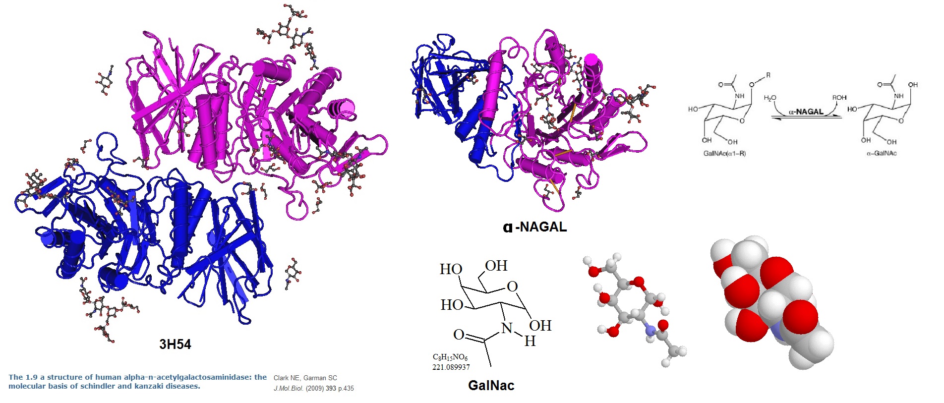
Figure 2: Molecular models of α-N-acetylgalactosaminidase (α-NAGAL, EC. 3.2.1.49). A lysosomal exoglycosidase that cleaves terminal α-N-acetylgalactosamine residues from glycopeptides and glycolipids. In humans, a deficiency of α-NAGAL activity results in the lysosomal storage disorders Schindler and Kanzaki diseases. The reaction catalyzed by the enzyme is shown in the right upper right. Molecular models for GalNAc are shown to the right. Models of the enzyme structure are shown on the left and in the upper middle panel. Ribbon diagrams of the human α-NAGAL dimer and monomer with the enzymatic product α-GalNAc in the active sites are shown (Clark and Garman, 2009).
Functions of O-GalNAc glycans
O-GalNAc glycosylation is a post-translational modification. O-GalNAc glycosylation appears to be an essential process since all mammalian cell types studied so far express polypeptide N-acetylgalactosamine (GalNAc) transferases (ppGalNAcTs). The enzyme polypeptide N-acetylgalactosamine (GalNAc) transferase (ppGalNAcT) when active initiates the covalent linkage of GalNAc to serine and threonine residues of proteins. Many ppGalNAcTs operate within multicellular organisms. However, they differ in expression patterns and substrate selectivity. It appears that ppGalNAcTs are important for differentially modulated regulatory processes in animal development, physiology, and possibly in disease. For example, animals lacking ppGalNAcT-1 are markedly impaired in immunoglobulin G production, have increased germinal center B-cell apoptosis and reduced levels of plasma B cells.
O-glycans are hydrophilic and usually negatively charged. These characteristics allow them to bind water and salts making them major contributors to the viscosity and adhesiveness of mucus. Removal of microbes and particles trapped in mucus is an important physiological process. O-GalNAC glycans significantly influence the conformation of the attached protein. O-glycosylation of mucins provides almost complete protection from protease degradation. Similarly, this can be the case for other glycoproteins as well. Furthermore, it is thought that O-glycans of cell-surface receptors may regulate receptor stability and expression levels. Also, selectin-glycan interactions are important for the attachment of leukocytes to the capillary endothelium during homing of lymphocytes or the extravasation of leukocytes during the inflammatory response. To exemplify this, the removal of core 2 O-GalNAc glycans from mice by eliminating the C2GnT-1 gene resulted in a sever deficiency in the immune system of these mice. Cancer cells that often express sialyl LewisX epitopes appear to use the selectin-binding properties of the glycan to invade other cell tissue. In cancer, it was observed that the biosynthesis of O-GalNAc glycans is often abnormal, which may affect the biology and survival of the cancer cell. Finally, O-glycosylated glycoproteins may be important during reproduction and fertilization.
References
Nathaniel E. Clark and Scott C. Garman; The 1.9 Å structure of human α-N-acetylgalactosaminidase: The molecular basis of Schindler and Kanzaki diseases. J Mol Biol. 2009 October 23; 393(2): 435–447. doi:10.1016/j.jmb.2009.08.021.
Delta masses: http://www.ionsource.com/; https://www.abrf.org/index.cfm/dm.home?AvgMass=all
Donald M. Marcus, Elvin A. Kabat, Gerald Schiffman (1964). "Immunochemical Studies on Blood Groups. XXXI. Destruction of Blood Group A Activity by an Enzyme from Clostridium tertium Which Deacetylates N-Acetylgalactosamine in Intact Blood Group Substances". Biochemistry 3: 437–443. doi:10.1021/bi00891a023
Tenno M, Ohtsubo K, Hagen FK, et al. Initiation of Protein O Glycosylation by the Polypeptide GalNAcT-1 in Vascular Biology and Humoral Immunity . Molecular and Cellular Biology. 2007;27(24):8783-8796. doi:10.1128/MCB.01204-07. http://www.ncbi.nlm.nih.gov/pubmed/17923703
NIH Book
Essentials of Glycobiology. 2nd edition. http://www.ncbi.nlm.nih.gov/books/NBK1896/ . Essentials of Glycobiology, 2nd edition. Editors: Ajit Varki, Richard D Cummings, Jeffrey D Esko, Hudson H Freeze, Pamela Stanley, Carolyn R Bertozzi, Gerald W Hart, and Marilynn E Etzler. Cold Spring Harbor (NY): Cold Spring Harbor Laboratory Press; 2009. ISBN-13: 9780879697709.