BNA based FISH probes
Recent advancements in the design and development of molecular probes and image analysis has made fluorescence in situ hybridization (FISH) a powerful tool. Although being a useful technique FISH is a fairly time-consuming procedure with limitations in sensitivity. Probes that exhibit higher DNA affinities promise to potentially improve the sensitivity of the technique. Artificial nucleotides such as bridged nucleic acids (LNAs) have been described for the development of chimeric LNA/DNA oligonucleotides as probes for fluorescence in situ hybridization on metaphase chromosomes and interphase nuclei.
Bridged nucleic acids (BNA3) are artificial bicyclic oligonucleotides that contain a five-membered or six-membered bridged structure with a “fixed” C3’-endo sugar puckering (Saenger 1984). The bridge is synthetically incorporated at the 2’, 4’-position of the ribose to afford a 2’, 4’-BNA monomer. The monomers can be incorporated into oligonucleotide polymeric structures using standard phosphoamidite chemistry. BNAs are structurally rigid oligo-nucleotides with increased binding affinities and stability. Oligonucleotide modifications are characterized by the presence of one or more bicyclic ribose analogs. The structural similarity to native nucleic acids and the presence of a nitrogen atom within the bicyclic ring leads to very good solubility in water and allows for easy handling of synthetic primers and probes. In contrast to peptide nucleic acids (PNAs) and minor groove binders (MGBs), but similar to LNAs, BNA monomers can be used for both primers and probes in real time quantitative polymerase chain reaction (RT-Q-PCR) assays. Compared to locked nucleic acids (LNAs) the substitution of DNA monomers with BNA monomers in oligonucleotides adds exceptional biological stability, resistance to nucleases and a significantly increased affinity to their complementary DNA targets.
Silahtaroglu et al. in 2003 reported results for a study where LNA substituted oligonucleotides of either the 23-bp human satellite-2 repeat sequence (ATT CCA TTC GAT TCC ATT CGA TC) or the 24-bp sequence composed of four blocks of the 6-bp telomere repeat (TTAGGG) have been used. The different LNA designs of the chimeric LNA/DNA probes for the human satellite–2 repeat used here are listed in table I. The desigin of a BNA based FISH probe is shown in table II.
Table I. The LNA/DNA mixmer FISH probes for human satellite-2 repeat sequence used in the study*.
*LNA substitutions are depicted in capital letters
Table II. The BNA/DNA mixmer FISH probes for human satellite-2 repeat sequence#.
|
BNA probe
|
ATtCcAtTcaGaTtCcAtTcGaTc
|
11
|
#BNA substitutions are depicted in capital letters.
For the telomere specific LNA probe, only the LNA-2 design, with an LNA substitution at every second nucleotide position, was synthesized together with a DNA control. Oligonucleotide FISH probes with different LNA substitution patterns, labels and hybridization conditions were used in a comparative study and subsequently the optimal conditions were determined for an efficient LNA-FISH protocol. All LNA-containing oligonucleotides for human satellite-2 and the telomere repeats gave prominent signals when used as FISH probes. For the human satellite-2 sequence, the LNA-2 design gave the best hybridisation results in the experiments performed. The LNA-3 probe, with every third oligonucleotide substituted with LNA, also gave hybridisation signals, albeit weaker than those obtained with the LNA-2 probes.
Repetitive elements comprise ~45% of the human genome and consist of interspersed repeats derived from non-autonomous or autonomous transposable elements and tandem repeats of simple sequences (satellite DNA) or complex sequences. The most abundant short interspersed nucleotide element (SINE) in human DNA is the Alu repeat, an ~282 bp non-LTR (Long Terminal Repeat) DNA sequence, which comprises 10% of the human genome and is present in ~1 million copies per haploid genome. Other abundant non-LTR sequences are long interspersed nucleotide elements (LINEs) of up to 6 kb that comprise ~20% of the human genome. LINE-1 elements are present at over 500 000 copies in the human genome. Only 3000–4000 are full length and 30–100 are active retrotransposons.
LINE-1 elements are usually methylated in somatic tissues, and LINE-1 hypomethylation is a common characteristic of human cancers. Alu sequences are also normally methylated in somatic tissues and are thought to become hypomethylated in human cancer cells. Not all Alus are hypomethylated in human cancers. Alu sequences located upstream of the CDKN2A promoter were found to be hypermethylated in cancer cell lines, and an Alu sequence located in intron 6 of TP53 showed extensive methylation in normal and cancer cells. LINEs and SINEs are interspersed throughout the genome, whereas satellite DNA is largely confined to the centromeres or centromere-adjacent (juxtacentromeric) heterochromatin and to the large region of heterochromatin on the long arm of the Y chromosome. Satellite a (Sata) repeats are composed of 170 bp DNA sequences and represent the main DNA component of every human centromere. Satellite 2 (Sat2) DNA sequences are found predominantly in juxtacentromeric heterochromatin of certain human chromosomes and are most abundant in the long juxtacentromeric heterochromatin region of chromosome (Chr) 1. Sat2 sequences are composed of variants of two tandem repeats of ATTCCATTCG followed by one or two copies of ATG. Both Chr1 Sata and Chr1 Sat2 sequences, as well as Sata repeats present throughout all the centromeres, are highly methylated in normal postnatal tissues, hypomethylated in sperm and often hypomethylated in various cancers (26–29). In addition, Sat2 sequences on Chr1 and Chr16 are also hypomethylated in the ICF (immunodeficiency, centromeric region instability and facial abnormalities) syndrome, which usually involves mutations in DNMT3B.
Probe design and preparation
The 23-bp human satellite-2 repeat sequence, attccattcgattccattcgatc, or a 24-bp telomere sequence (ttagggttagggttagggttaggg) representing 4 blocks of 6-bp telomere repeat (ttaggg) are used for the BNA/DNA mixmers with different BNA substitution patterns (Table I). All mixmers oligonucleotides are both synthesized either with a Cy3 or a biotin group at the 5’ end. An unsubstituted DNA oligonucleotide probe is used as a control in each experiment. All the oligonucleotide probes are kept frozen until used in aliquots of distilled water.
Fluorescence in situ hybridization
FISH is carried out as described previously (Silahtaroglu et al., 1998) with the following modifications. The amount of probe is 6.4, 10, 13.4 and 20 pmoles. Denaturation of the target DNA and the probe are performed at 75oC for 5 minutes either separately using 70% formamide or simultaneously under the coverslip in the presence of hybridization mixture containing 50% formamide. In addition, the effect of denaturation is also tested. Two alternative hybridization mixtures are used: 50% formamide/2xSSC (pH 7.0) /10% dextran sulphate or 2xSSC (pH 7.0) /10% dextran sulphate. Hybridization times include 30 min, 1 hr, 2 hrs, 3 hrs and overnight. Hybridization temperatures include: 37°C, 55°C, 60°C and 72°C. Post-washing is either as for standard FISH, or with 50% formamide/2xSSC at 60°C, or without formamide. Hybridization signals with biotin labeled LNA/DNA mixmers are visualized indirectly using two layers of fluorescein labeled avidin (Vector Laboratories, USA) linked by a biotinylated anti-avidin molecule, which amplifies the signal 8-64 times. The hybridization of Cy3-labeled molecules is visualized directly after a short washing procedure. Slides are mounted in Vectashield (Vector Laboratories, USA) containing 4´-6´-diamidino-2-phenylindole (DAPI). The whole procedure is carried out in the dark. The signals are visualized using a Leica DMRB epifluorescence microscope equipped with a SenSys charge-coupled device camera (Photometrics, Tucson, AZ, USA), and IPLAB Spectrum Quips FISH software (Applied Imaging international Ltd., Newcastle, UK) within two days after hybridization. 20 metaphases are analyzed after each hybridization experiment.
Repetitive element in the human genome
Description, primers and probes of repetitive elements
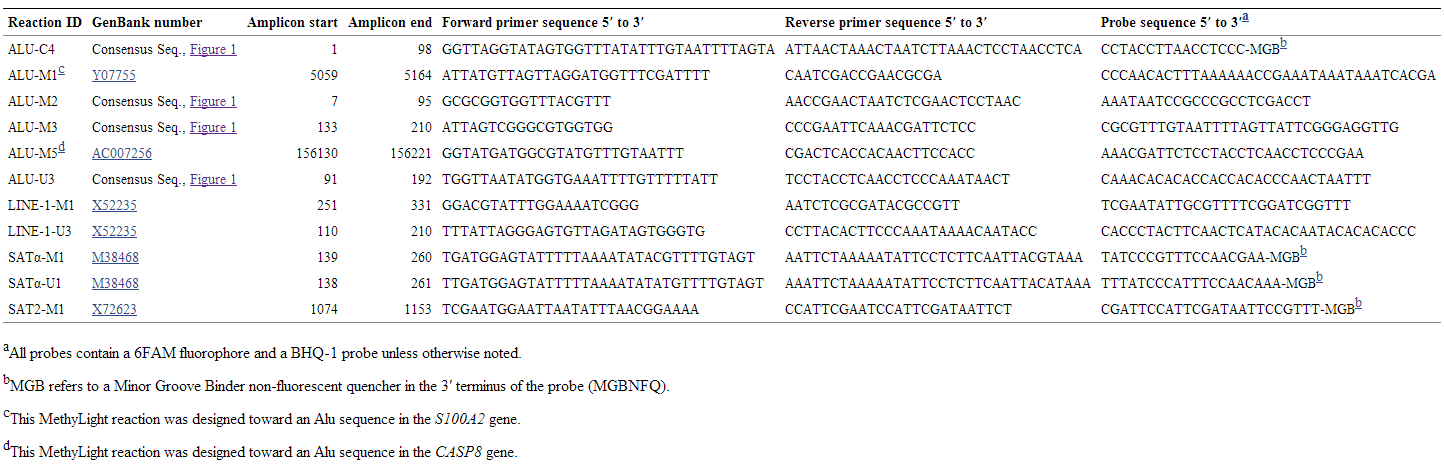
(Source: Weisenberger et al. 2005)
MethyLight
MethyLight is a sodium-bisulfite-dependent, quantitative, fluorescence-based, real-time PCR method to detect and quantify DNA methylation in genomic DNA with high sensitivity. The technique relies on methylation-specific priming combined with methylation-specific fluorescent probing. The combination of the two methylation-specific detection principles results in a highly methylation-specific detection technology. It allows to detect very low frequencies of hypermethylated alleles. The high sensitivity and specificity of MethyLight make it uniquely well suited for detection of low-frequency DNA methylation biomarkers. DNA and RNA based biomarker such as cell-free nucleic acids, mRNA and microRNA releases into the blood stream of cancer patients are increasingly used as target molecules to detect different tumor types or diseases. The quantitative accuracy of real-time PCR and the flexibility to design bisulfite-dependent, methylation-independent control reactions allows for a quantitative assessment of low-frequency methylation events.
References
Jeanpierre, M. (1994). Human satellites 2 and 3. Annals of Genetics 37, 163-171.
Moyzis RK, Buckingham JM, Cram LS, Dani M, Deaven LL, Jones MD, Meyne J, Ratliff RL, Wu JR. (1988) A highly conserved repetitive DNA sequence, (TTAGGG)n, present at the telomeres of human chromosomes. Proc Natl Acad Sci U S A. 85(18):6622-6626.
Sanger Wolfram; Principles of Nucleic Acid Structure. Springer-Verlag, New York, Berlin, Heidelberg, London, Paris, 1984.
Silahtaroglu, A.N., Hacihanefioglu, S., Guven, G.S., Cenani, A., Wirth, J., Tommerup, N., Tumer, Z.(1998) Not para-, not peri-, but centric inversion of chromosome 12. Journal of Medical Genetics 35(8), 682-684.
Silahtaroglu, A.N., Tommerup, N., Vissing, H. (2003) FISHing with Locked Nucleic Acids (LNA): Evaluation of different LNA/DNA Mixmers. Molecular and Cellular Probes 17(4), 165-169.
Daniel J. Weisenberger, Mihaela Campan, Tiffany I. Long, Myungjin Kim, Christian Woods, Emerich Fiala, Melanie Ehrlich and Peter W. Laird; Analysis of repetitive element DNA methylation by MethyLight. Nucleic Acids Research, 2005, Vol. 33, No. 21 6823–6836. doi:10.1093/nar/gki987.